Multiparameter References
Navigation
Capnography | Electrocardiography | | Blood Pressure
Capnography
Normal | Cardiac Oscillations | Hypercapnia Rebreathing | Airway Obstruction
Decreasing ETCO2
End-tidal CO2 is determined by three physiological components:
1. Metabolic Rate
2. Circulation
3. Alveolar VentilationIf 2 of these are stable, capnography tells you about the unstable oneWhat is Normal and AbnormalHypocapnia <35mmHg
Normcapnia 35-45mmHg
Hypercapnia >45mmH
Causes of Increased ETCO2
Hypoventilation1. Anaesthetic depth is too deep
2. Is the patient obese, pregnant or poorly positioned
3. Increased dead spaceIncreased production of CO21. Metabolic rate too high due to increased temperature, shivering, seizures, painIncreased inspired CO2 concentration (Rebreathing)1. O2 flow rate too little - increase free gas flow
2. Replace old soda lime
3. One way valve broken
4. May be seen due to insufflation during laproscopic surgery
Causes of Decreased ETCO2
Hyperventilation1. Anaesthetic depth too light
2. Severe hypoxaemiaReduced CO2 Production1. Hypothermia - lack of active heating
2. Hypometabolic state such as hypothyroidismIncreased Alveolar Dead Space1. Hypotension due to low cardiac output
2. Pulmonary embolism e.g. air, thromboembolism
3. High inspiratory pressure during IPPVSudden Decrease in ETCO21. Equipment calibration, blockage, disconnection or displacement
2. Oesophageal intubation
3. Ventilation failure - apnea or respiratory arrest
4. Circulation failureSampling Errors
Normal Capnograph

1. The wave should roughly be square or rectangular
2. The base should be 0mmHg on inspiration
3. Rises steeply from 0 to the plateua and then drops steeply again
Cardiac Oscillations - A variation of normal
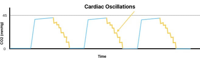
1. The wiggle on the downward aspect matches heart rate
2. Normally seen in patients with a slow respiratory rate and or barrel chested breeds
Hypercapnia - Appears normal but plateau is elevated above normal range
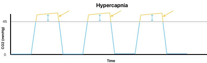
💡 Causes1. Hypoventilation
2. Increased CO2 production from metabolism
3. Increased cardiac output
Rebreathing - Looks normal except the base does not touch the baseline
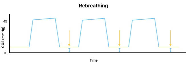
💡 Causes - Patient is rebreathing CO21. Excess dead space for patient (tube too long possibly)
2. Flow rate is too low
3. Soda lime needs replacing
4. One way valve not working
Airway Obstruction - ‘Shark fin’
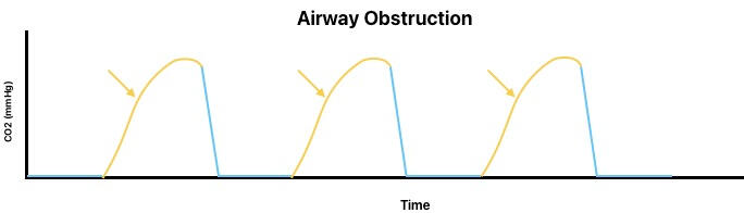
💡 Causes - Increased expiratory airway resistance1. Bronchospasm
2. Mucous obstructing tube lumen
3. Kinked tube
4. ET tube too far in and open end is against tracheal wall
Decreasing ETCO2
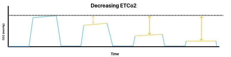
The plateau end tital CO2 is decreasing💡 Causes for gradual decrease1. Hyperventilation
2. Leaking cuff💡 Causes for a rapid change1. Disconnected circuit
2. Pulmonary embolism
3. Decreased cardiac output, which can be a sign of impending cardiac arrest